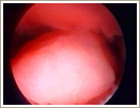
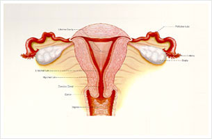
Hysteroscopy is a procedure carried out to view the inside of the uterus. The instrument used looks like a thin telescope. It is introduced into the vagina, through the cervix , slowly moves through the cervical canal into the uterine cavity.
View through a hysteroscope
A liquid is usually used to distend the uterine cavity to enable proper inspection. The video below is typical of a normal uterus during hysteroscopy. This is obtained by attaching a light weight camera to the hysteroscope.
This procedure can be done in an office setting. There is some degree of discomfort but most women find it bearable. However, some patients do prefer to be sedated or even have a general anesthesia.
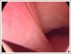
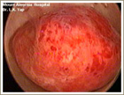
At different time during the cycle the uterine cavitylooks different. During the period there is much shedding of the lining and the view is poor.
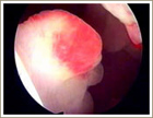
View of uterine cavity during menses
Uses
The hysteroscope is useful in the investigation and treatment of:
- Abnormal uterine bleeding, including heavy, irregular, prolonged and inter-menstrual bleeding
- Infertility
- Miscellaneous
Abnormal Uterine Bleeding:
Some common local causes of abnormal bleeding are:
- Polyps, both cervical and endometrial
- Fibroids
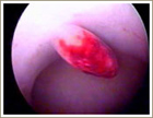
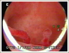
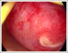
Polyps - Cervical
Intracavitary Fibroid
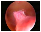
A simple polyp arising from the cervical canal.
Intracavitary Fibroid
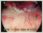
This cervical polyp has a long stalk.
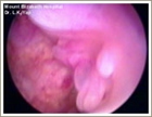
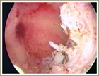
Endometrial Polyps
Small polyp arising from endometrium.
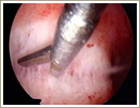
Scissore excision of polyp being done.
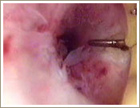
A simple endometrial polyp with a stalk arising from a small base.
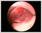
On first look it appeared to be a cervical polyp but a closer look shows that it arises from the uterine cavity.
Multiple endometrial polyps commonly occur and are often responsible for bleeding persisting after D & C. Note the redness at the tip, often seen in polyps with a tendency to bleed.
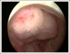
Sessile Polyps: Some polyps have a broad base and appear flattened.
On inspection it is often difficult to tell if the lesion is a polyp or fibroid.
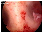
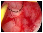
Uterine Fibroids
Various Pathologies seen on Hysteroscopy
Uterine Septum
A simple polyp arising from the cervical canal.
Endometrial Hyperplasia
This cervical polyp has a long stalk.
Endometrial Cancer
Miscellaneous
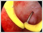
Forgotten IUCD Causing bleeding.
Operative Hysteroscopy
Hysteroscopes are extremely useful for diagnosing problems.
With special attachments fitted the instrument is extremely versatile and can in the right hands be used for removing the problems. Operative work includes:
- Polypectomy – uterine polyp removal
- Myomectomy – removal of fibroids in the uterine cavity
- Resection of septum
- Endometrial ablation
- Sterilisation
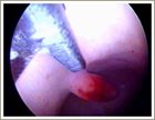
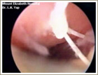
Polypectomy
Inspection of a small endometrial polyp.
Resection of polyp ensures thet recurrence at the same site is unlikely.
Myomectomy
Small fibroid may not cause problems but can cause difficulty in implantation of the embryo and during pregnancy can grow to a size large enough to cause problems like bleeding and possibly early miscarriage.
Some fibroids are small enough to be avulsed blindly as done in the past but often a stump is left behind. Removal with the loop is useful in preventing recurrence.
Large fibroids in the cavity can be removed by resecting . Small pieces are taken during each sweep with the loop and the movement is continued till the whole fibroid is removed.
In this video most of the fibroid has been removed leaving only about 20 percent of the original fibroid. This remainder too is removed to prevent recurrence.
Resection of septum